MRI Cauda Equina: Complete Guide to Emergency Imaging, Timing, and What to Expect

Introduction
Magnetic resonance imaging (MRI) serves as the gold standard diagnostic tool for suspected cauda equina syndrome, with urgent mri scanning required within 4 hours of clinical suspicion to prevent permanent neurological damage. Emergency mri can identify cauda equina compression at the lumbar region and differentiate between cases requiring emergency surgical decompression and non-urgent conditions affecting the spinal canal.
Despite only 4.7% of suspected cauda equina syndrome cases requiring emergency surgery, delayed diagnosis can result in permanent urinary incontinence, bowel dysfunction, and sexual dysfunction, making timely imaging a critical medicolegal and patient safety priority. Early recognition of CES is critical, as delayed treatment can lead to permanent neurological damage. The majority of patients with CES who undergo surgery within 24 hours of symptom onset have better outcomes compared to those who are treated later. However, there is debate regarding the exact timing of surgical intervention for cauda equina syndrome, with some studies suggesting no significant difference in outcomes between surgeries performed within 24 hours and those performed later.
What This Guide Covers
This comprehensive resource outlines emergency mri protocols for suspected ces, national timing standards requiring hours magnetic resonance imaging availability, scan interpretation guidelines, and post-scan management pathways. We cover red flag symptoms recognition, urgent decompression decisions, and transfer protocols, but do not address routine spinal mri procedures or non-emergency diagnostic imaging. The lack of timely access to MRI for suspected CES is a significant factor contributing to delays in diagnosis and treatment.
Who This Is For?
This guide is designed for emergency department staff, consultant spinal surgeons, general practitioners, and healthcare professionals managing spinal emergencies. Whether you’re evaluating patients presenting with bilateral sciatica and saddle anaesthesia or coordinating suspected ces referrals between spinal units, you’ll find evidence-based protocols for emergency mri provision.
Why This Matters?
Understanding cauda equina syndrome diagnosis prevents permanent neurological complications that affect bladder function, sexual function, and lower limbs mobility. The healthcare safety investigation branch emphasizes that prompt diagnosis through emergency mri scan reduces medicolegal risks and ensures optimal patient outcomes, even when 95% of scans return negative results. Patients with suspected CES often experience delays in receiving MRI scans, which can lead to prolonged suffering and increased risk of permanent damage. The timing of surgical decompression is critical in cauda equina syndrome, with evidence suggesting that earlier intervention leads to better outcomes. The rising demand for MRI scans for suspected CES is driven by increased awareness of the condition and the potential for litigation due to missed diagnoses. Claims related to delayed management of CES average around £400,000, with some organizations reporting even higher averages per case.
What You’ll Learn:
- When emergency mri is required for suspected cauda equina syndrome
- National timing standards and 24/7 mri provision requirements
- How to interpret mri findings and understand positive mri vs negative results
- Transfer protocols and surgical intervention pathways following diagnostic imaging
- How MRI allows doctors to plan appropriate and timely treatment to prevent irreversible damage such as permanent paralysis or loss of bladder and bowel control
Anatomy and Physiology of the Cauda Equina
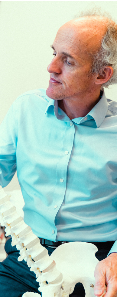
The cauda equina is a critical structure located at the lower end of the spinal cord, within the spinal canal. It consists of a bundle of lumbar and sacral nerve roots that resemble a horse’s tail—hence the name “cauda equina.” These nerve roots are responsible for transmitting signals that control movement and sensation in the lower limbs, as well as vital functions such as bladder and bowel control.
In cauda equina syndrome (CES), these nerve roots become compressed or damaged, leading to a range of neurological symptoms. Because the cauda equina sits within the protective spinal canal, any condition that narrows this space—such as a herniated disc, spinal stenosis, or tumor—can put pressure on the nerve roots. Understanding the anatomy and physiology of the cauda equina is essential for recognizing the signs of cauda equina syndrome CES and ensuring prompt, effective intervention to prevent permanent disability.
Causes of Cauda Equina Syndrome
Cauda equina syndrome can develop from a variety of underlying conditions that result in compression or injury to the cauda equina nerve roots. The most common cause is lumbar disc herniation, where a herniated disc in the lower back presses on the nerve roots. Other frequent causes include spinal stenosis (narrowing of the spinal canal), metastatic spinal cord compression from cancer, and spinal tumors. Less commonly, traumatic injuries, infections, or inflammatory diseases can also lead to CES.
Regardless of the cause, the result is often severe pain, numbness, weakness, and bowel dysfunction due to impaired nerve signaling. In some cases, cauda equina syndrome may occur suddenly and without warning, making prompt diagnosis and intervention crucial to prevent irreversible damage to the spinal cord and cauda equina nerve roots.
Understanding MRI in Cauda Equina Syndrome
Magnetic resonance imaging mri provides superior soft tissue visualization of cauda equina nerve roots within the spinal canal, enabling precise identification of nerve root compression from lumbar disc herniation, spinal tumours, or metastatic spinal cord compression. The most common cause of CES is lumbar disc herniation, particularly at the L4-L5 and L5-S1 levels.
Unlike CT scans or plain radiographs, mri scan technology delivers detailed images of herniated disc material, spinal stenosis, and cauda equina compression that determine whether emergency surgical decompression is necessary. This imaging superiority becomes critical when evaluating the rare but serious condition affecting approximately 1 in 100,000 individuals annually. Cauda Equina Syndrome (CES) is a rare condition characterized by dysfunction of the sacral and lumbar nerve roots in the vertebral canal. Massive central lumbar disc herniation, spinal stenosis, spinal tumors, spinal infections, trauma, and inflammatory conditions are conditions that can cause cauda equina syndrome. Spinal stenosis involves narrowing of the spinal column, which can compress nerve tissues and lead to symptoms such as pain, numbness, and weakness.
The cauda equina nerve roots extending below the spinal cord termination control essential functions including bladder function, bowel control, and sensation to the pelvis and lower limbs, making accurate imaging interpretation vital for preventing permanent disability.
MRI vs Other Imaging Methods
Emergency mri provides unparalleled soft tissue contrast compared to CT myelography, revealing disc herniation details and spinal nerve compression with superior accuracy. CT myelography serves as an alternative only when mri findings are inconclusive or patients have absolute contraindications including certain pacemakers or severe claustrophobia requiring emergency department sedation protocols. The lack of 24/7 MRI availability in many hospitals contributes to delays in diagnosing cauda equina syndrome, which is critical for timely intervention. Additionally, MRI can identify potential mimics of cauda equina syndrome, such as aortic dissection or spinal infarction. Abdominal pain should also be considered in the differential diagnosis, as it may be related to or mistaken for symptoms of cauda equina syndrome or other underlying conditions.
This connects to emergency mri protocols because alternative imaging methods introduce diagnostic delays that can worsen neurological symptoms and compromise surgical outcomes for patients with confirmed cauda equina compression.
Types of MRI Findings in CES
Positive mri findings include central lumbar disc herniation causing >75% spinal canal occlusion, disc sequestration compressing cauda equina nerve roots, lumbar spinal stenosis with significant nerve compression, and spinal tumours affecting the lumbar region. MRI can also reveal pathology involving the vertebral bodies, such as fractures, compressions, or infections like spondylodiscitis, which may contribute to nerve compression and the development of cauda equina syndrome. These findings directly indicate need for urgent decompression and immediate consultant spinal surgeon consultation. Patients with incomplete cauda equina syndrome should be decompressed immediately to improve neurologic and urologic outcomes.
Building on imaging superiority, mri findings guide surgical intervention decisions within hours of symptom onset, distinguishing surgical emergencies from conditions causing bilateral leg pain without cauda equina involvement.
Transition: Understanding these imaging capabilities explains why national guidance mandates specific timing standards for emergency mri access in suspected ces cases.
When Emergency MRI is Required for Suspected CES
Clinical suspicion of cauda equina syndrome secondary to nerve root compression demands immediate diagnostic imaging to prevent irreversible neurological damage affecting urinary flow, bowel control, and sexual function.
Red Flag Symptoms Requiring Urgent MRI
Patients presenting with bilateral sciatica affecting both legs, saddle anaesthesia (numbness around the buttocks and perineal area), urinary retention or urinary incontinence, and new-onset bowel dysfunction require emergency mri scan within 4 hours. Additional concerning neurological symptoms include severe pain radiating to lower limbs, progressive weakness affecting pelvis and lower limbs, and altered sensation in the lumbar region distribution. CES can lead to impairment of bladder, bowel, or sexual function, as well as perianal or saddle numbness.
Saddle anaesthesia represents numbness or altered sensation in the “saddle” area where you would sit on a horse – the buttocks, inner thighs, back of legs, and genital region, indicating potential cauda equina nerve root involvement.
Timing Standards and Guidelines
National treatment guidelines mandate urgent mri within 4 hours of clinical suspicion, with mri findings reported within 1 hour by consultant radiologists or appropriately trained specialists. The british association of spinal surgeons emphasizes 24/7 emergency mri availability at hospitals accepting spinal services referrals, ensuring patients referred for suspected ces receive timely diagnosis regardless of presentation time.
Unlike routine spinal imaging that can be scheduled during normal hours, suspected cauda equina syndrome requires immediate mri provision comparable to stroke or heart attack protocols.
Clinical Decision Making
Emergency department physicians maintain a low threshold for emergency mri given the potential for permanent disability and significant medicolegal implications of delayed diagnosis. The Society of British Neurological Surgeons and the British Association of Spine Surgeons recommend a low threshold for requesting an emergency MRI scan for suspected cauda equina syndrome. Clinical diagnosis alone cannot reliably distinguish surgical emergencies from non-urgent back pain conditions, making diagnostic imaging essential even when clinical symptoms appear mild initially.
Key Points:
- 95% of suspected ces cases have negative mri results, but urgent scanning remains essential
- 4-hour window from clinical suspicion to mri scan acquisition represents the critical target
- 24/7 mri provision is mandatory at hospitals accepting emergency spinal referrals
Transition: These timing requirements necessitate specific emergency mri protocols that differ significantly from routine spinal imaging procedures.
The MRI Process for Cauda Equina Syndrome
Emergency mri protocols for suspected cauda equina syndrome require coordinated efforts between emergency department staff, radiology services, and spinal units to achieve national guidance timing standards while ensuring patient safety throughout the diagnostic process.
Step-by-Step: Emergency MRI Protocol
When to use this: For emergency department and referring clinicians managing patients with suspected ces symptoms requiring urgent evaluation.
- Clinical Assessment: Emergency department senior clinician completes red flag symptoms evaluation within first hour, documenting bilateral leg pain, saddle anaesthesia, urinary symptoms, and bowel dysfunction using standardized assessment tools.
- Urgent MRI Request: Direct communication between emergency department and consultant radiologist or specialist trainee, bypassing routine referral pathways to ensure immediate scan scheduling and appropriate clinical priority.
- Patient Preparation: Rapid contraindication screening for metallic implants, pacemakers, or pregnancy, with claustrophobic patients receiving appropriate sedation protocols to prevent diagnostic delays from incomplete scanning.
- Lumbar Spine MRI: Comprehensive lumbar region imaging typically requiring 21-minute scan duration, focusing on spinal canal assessment from L1-S1 levels to identify cauda equina compression sources.
- Immediate Reporting: Consultant radiologist or specialist trainee provides preliminary interpretation within 1 hour, with positive mri findings triggering immediate consultant spinal surgeon contact for emergency surgery evaluation.
Comparison: In-Hours vs Out-of-Hours MRI
Feature | In-Hours Provision | Out-of-Hours Provision |
|---|---|---|
Reporting Time | Immediate consultant availability | Specialist trainee with 24-hour consultant verification |
Transfer Requirements | Local hospital management | Potential transfer to tertiary spinal services |
Surgical Decision | Same-day consultant spinal surgeon consultation | Emergency surgery coordination through regional networks |
Only 14% of UK trusts currently provide comprehensive out-of-hours emergency mri, necessitating patient transfer protocols to tertiary centers with 24/7 spinal services capability when time-critical diagnosis cannot be achieved locally. The National Institute for Health and Care Excellence recommends that MRI should be available at the referring hospital 24 hours a day, 7 days a week for suspected cauda equina syndrome. Access to MRI for suspected cauda equina syndrome (CES) is often limited to major trauma centers and neurosurgical units, creating challenges for patients in regional hospitals. The cost of CES-related litigation claims for the NHS was reported to be £68 million for the years 2014/15 to 2015/16.
Transition: Despite robust protocols, several systematic challenges affect emergency mri provision for suspected cauda equina syndrome across healthcare systems.
Common Challenges and Solutions
Healthcare systems face significant obstacles in providing optimal emergency mri access for suspected ces, requiring coordinated solutions that balance cost-effectiveness with patient safety priorities and medicolegal risk management.
Challenge 1: Limited Out-of-Hours MRI Availability
Solution: Establish formal transfer protocols to tertiary centers with 24/7 mri provision and regional spinal network coordination, ensuring patients referred for suspected ces receive timely diagnosis regardless of referring hospital capabilities.
Current data shows only 14% of UK trusts provide comprehensive out-of-hours emergency mri, creating geographic disparities in access that require systematic network solutions rather than individual hospital investments.
Challenge 2: High Negative Scan Rates (95%+)
Solution: Maintain low clinical thresholds for emergency mri despite high negative rates, emphasizing that cost considerations cannot override patient safety and medicolegal protection when permanent neurological damage represents the alternative outcome.
The healthcare safety investigation branch reinforces that false negative consequences (missed cauda equina syndrome) far outweigh resource utilization concerns given the irreversible nature of untreated nerve compression.
Challenge 3: Reporting and Transfer Delays
Solution: Implement streamlined reporting protocols with specialist trainee out-of-hours coverage, consultant verification systems within 24 hours, and direct communication pathways between diagnostic imaging and spinal surgery services.
Studies indicate 50% of negative emergency mri cases experience discharge delays exceeding 24 hours due to reporting and communication inefficiencies rather than clinical necessity.
Transition: Addressing these challenges requires systematic implementation of evidence-based protocols that prioritize patient outcomes while maintaining sustainable healthcare delivery models.
Complications of Cauda Equina Syndrome
When cauda equina syndrome is not diagnosed or treated quickly, the consequences can be life-altering. Permanent nerve damage is a significant risk, leading to chronic pain, persistent numbness, and ongoing weakness in the lower limbs. Many patients experience urinary incontinence or urinary retention, as well as bowel dysfunction, which can severely impact daily life and independence. Sexual dysfunction is another common complication, further affecting quality of life.
Delayed diagnosis increases the likelihood of these complications, making it essential for healthcare providers to recognize the signs of cauda equina syndrome and act swiftly. Early intervention is the best way to minimize the risk of permanent disability and help patients regain as much function as possible.
Treatment Options for Cauda Equina Syndrome
The primary treatment for cauda equina syndrome is emergency surgical decompression, which aims to relieve pressure on the affected nerve roots as quickly as possible. Magnetic resonance imaging (MRI) is essential for confirming the diagnosis and guiding the surgical approach. The sooner surgical decompression is performed, the better the chances of preventing long-term complications and restoring normal function.
In addition to surgery, patients may benefit from supportive treatments such as pain management, physical therapy, and specialized rehabilitation for bladder and bowel function. Prompt diagnosis using magnetic resonance imaging mri, followed by timely surgical intervention, is critical for optimizing outcomes and reducing the risk of permanent nerve damage.
Patient Education and Support
For patients presenting with suspected cauda equina syndrome, education and support are vital components of care. Healthcare providers should clearly explain what cauda equina syndrome is, its potential causes, and the importance of recognizing red flag symptoms such as severe pain, numbness, and bowel dysfunction. Patients need to understand that prompt diagnosis and surgical intervention can make a significant difference in their recovery and long-term health.
It is also important to discuss the role of MRI in diagnosing CES and to set expectations about the urgency of treatment. Ongoing support, including access to rehabilitation services and counseling, can help patients manage any residual symptoms and adjust to changes in their daily lives. Empowering patients with knowledge about cauda equina syndrome and the importance of seeking immediate medical attention for red flag symptoms can improve outcomes and reduce the risk of delayed diagnosis.
Conclusion and Next Steps
Emergency mri remains the definitive diagnostic tool for suspected cauda equina syndrome, with strict timing standards essential for optimal patient outcomes despite the reality that 95% of urgent scans return negative results, reinforcing the critical balance between patient safety and resource management in emergency spinal care.
To get started:
- Recognize red flag symptoms requiring urgent mri within 4 hours of clinical suspicion
- Ensure 24/7 mri provision availability or establish transfer protocols to tertiary spinal services
- Implement rapid reporting systems with consultant verification for out-of-hours emergency mri scan results
Related Topics: Brief exploration of emergency surgical decompression techniques, regional spinal network development, and medicolegal considerations provides additional context for healthcare teams managing suspected ces and other spinal emergencies.
Additional Resources
- GIRFT 2023 cauda equina syndrome pathway guidelines and national treatment standards
- Royal College of Radiologists emergency mri provision requirements and timing benchmarks
- British Association of Spinal Surgeons red flag symptoms assessment tools and referral criteria
- Regional spinal network contact directories for emergency transfer coordination and consultant spinal surgeon consultation