Spine MRI Scan: Uses, Procedure, and Risks

A spine MRI scan uses magnetic fields and radio waves to diagnose spinal problems. If you’re in severe back pain or numbness, your doctor might order this scan. Here’s what you need to know.
Quick Facts
- A spine MRI scan is a non-invasive test that produces detailed images of the spine without radiation so it’s a safer option compared to other imaging tests.
- Spine MRI scans are necessary for diagnosing spine conditions like disc disease and spinal stenosis and plays a big role in early diagnosis and treatment planning.
- While generally safe, spine MRI scans have limitations like false positives and size restrictions for bigger patients; it should be used in conjunction with clinical exam for accurate diagnosis.
What is a Spine MRI Scan?
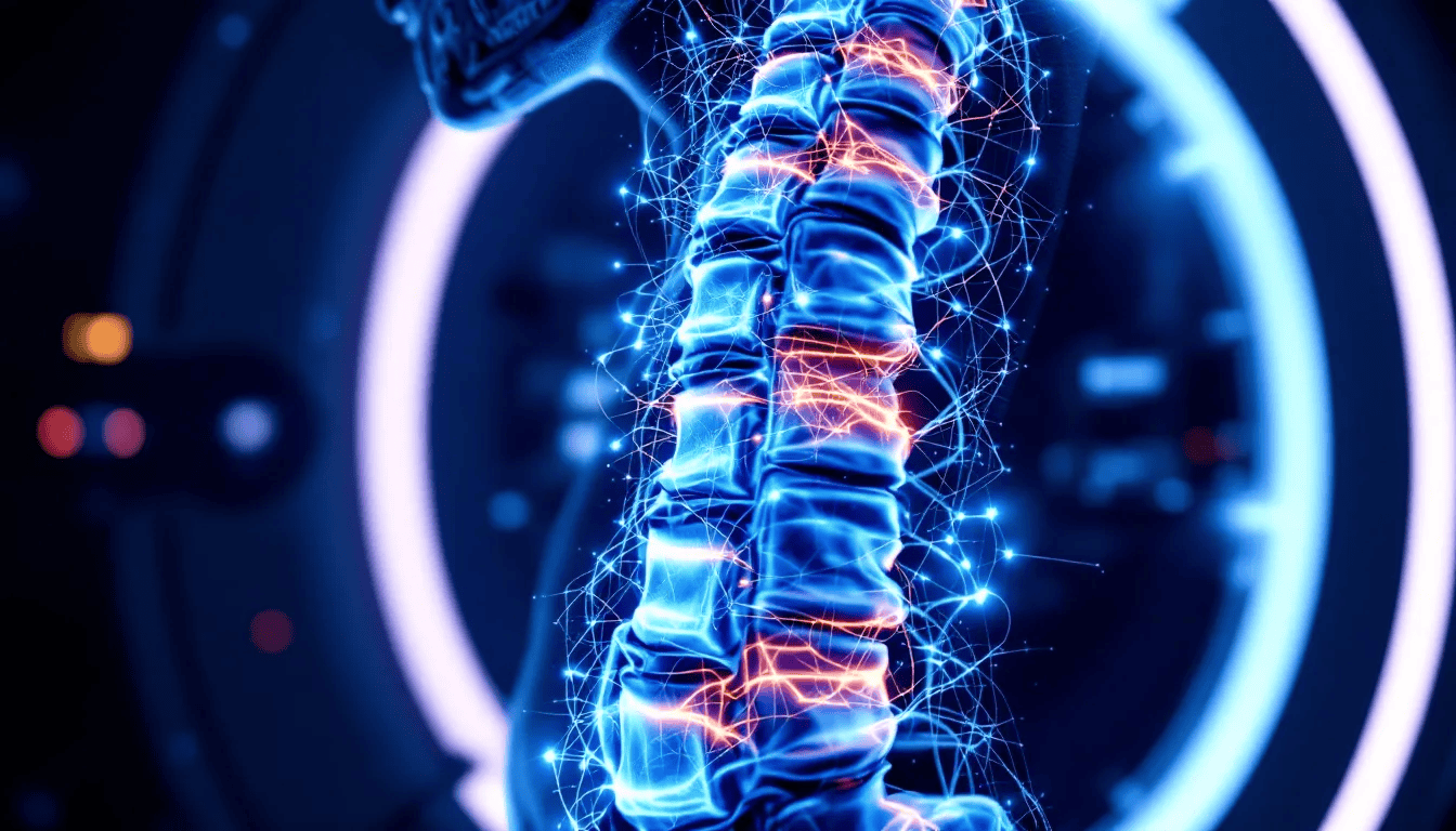
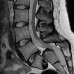
A spine MRI scan is a noninvasive diagnostic tool that uses a magnetic field and radiofrequency pulses to produce detailed images of the spinal column and surrounding soft tissues. Unlike X-rays and CT scans, an MRI scanner does not use radiation, making it a safer option for imaging tests. This advanced technology is capable of differentiating between diseased and normal tissues, providing highly sensitive and accurate assessments.
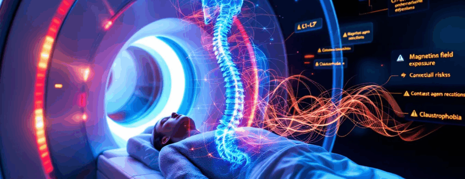
During the MRI procedure, patients lie on a table that slides into a cylindrical magnet, which generates the magnetic field necessary for imaging. Coils within the MRI unit send and receive radio waves that produce the signals required to create the images. The entire process typically lasts between 30 to 60 minutes, depending on the area being scanned, and patients are often given earplugs to minimize the noise from the MRI machine.
This detailed imaging capability is particularly useful for examining the spinal cord, lumbar spine, and spinal canal, offering insights that other imaging tests may not provide. The MRI machine’s ability to generate high-quality images without the use of radiation makes it an invaluable tool in modern medical diagnostics.
The Importance of Spine MRI Scans

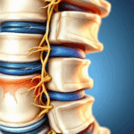
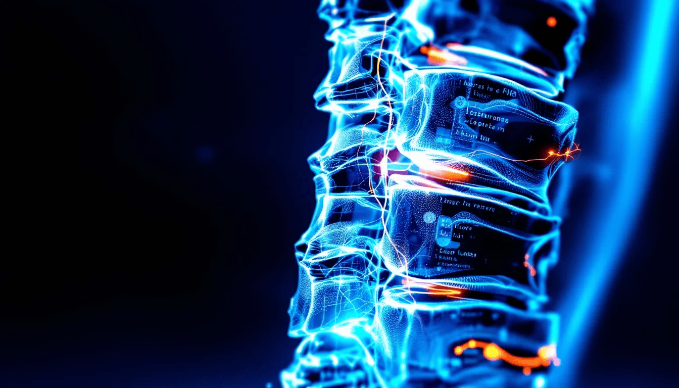
Early diagnosis of lumbar spine issues is vital for effective treatment. A lumbar spine MRI can identify various components such as bones, intervertebral discs, nerves, and surrounding soft tissues, providing a comprehensive view of the spinal column. This detailed imagery is crucial for pinpointing the exact cause of back pain and other spinal issues.
Spine MRI scans are indispensable in diagnosing conditions like spinal stenosis and assessing the overall health of the spinal canal. The high-quality images produced by magnetic resonance imaging (MRI) not only help in early diagnosis but also in planning appropriate treatment strategies. This makes MRI scans an essential tool in any comprehensive spinal health assessment.
Types of Spine MRI Scans
There are four primary types of spine MRI scans, each focusing on a different section of the spine: cervical, thoracic, lumbar, and sacral.
A cervical spine MRI scan concentrates on the neck area, covering vertebrae C1 to C7. This type of scan is crucial for diagnosing issues related to neck pain, nerve compression, and other conditions affecting the upper spine.
The thoracic spine MRI scan examines the mid-back region, specifically vertebrae T1 to T12. It is particularly useful for identifying problems related to the thoracic spinal column, such as herniated discs and spinal stenosis in the mid-back area.
The lumbar spine MRI scan targets the lower back, focusing on the last five vertebrae from L1 to L5. This type of scan is often requested for patients experiencing severe lower back pain, sciatica, or suspected lumbar disc herniation.
Each type of spine MRI scan provides unique insights that are essential for accurate diagnosis and effective treatment planning.
Conditions Detected by Spine MRI Scans
Spine MRI scans are essential tools for diagnosing a variety of conditions. For instance, they can identify pinched nerves, muscle weakness, and severe lower back pain. These scans excel in visualizing soft tissues, making them ideal for detecting spinal infections and tumors located in or around the spinal cord.
Conditions such as disc disease, nerve compression, and pinched nerves can be identified using MRI, which provides detailed images that guide treatment decisions. The clarity of MRI images ensures that doctors can pinpoint the exact cause of these conditions and develop effective treatment plans.
MRI scans also play a critical role in assessing trauma-related injuries to the spinal cord. They can identify spinal birth defects and abnormalities in alignment, providing a comprehensive view of the spine’s health. This makes MRI an invaluable tool for diagnosing a wide range of spinal conditions and ensuring timely and appropriate interventions.
When You Might Need a Spine MRI Scan
A doctor may suggest an MRI if you experience severe or persistent back pain, especially if it’s accompanied by neurological symptoms such as numbness or tingling. Warning signs for needing an MRI include loss of bladder or bowel control, difficulty walking, severe unexplained back pain, or weakness in the legs.
MRI can help confirm serious spinal pathologies that, if untreated, may lead to worse patient outcomes. If back pain does not improve after several weeks, an MRI may be necessary to investigate potential underlying issues.
If pain in and around the spine lasts more than a few weeks and interferes with daily life, seeking a spinal MRI scan is advisable.
Preparing for Your Spine MRI Scan
Preparing for a spine MRI scan involves a few crucial steps. First, inform the medical staff about any health problems, recent surgeries, allergies, or pregnancy before the scan. This information helps the medical team ensure your safety and the accuracy of the MRI images.
You will need to change into a hospital gown, remove any jewelry, and empty your pockets to avoid interference with the magnetic resonance imaging (MRI) machine. Generally, you can eat and drink before a spine MRI scan, unless advised otherwise by your doctor.
Following these preparation steps ensures a smooth and efficient MRI procedure.
What to Expect During a Spine MRI Scan
The duration of a spine MRI scan can vary, typically lasting between 15 to 90 minutes based on the area being examined and the number of images required. A lumbar MRI scan usually takes about 30 minutes but may take longer for specific details. During the procedure, patients are positioned on a moveable exam table, which may use straps and bolsters for better stability.
MRI machines emit loud tapping sounds due to the electric current cycling through the scanner coils. During the procedure, patients are often given earplugs. They may also receive headphones to help reduce noise.
MRI exams can be uncomfortable for some due to the need for patients to remain still in a confined space. Patients may feel a slight warmth in the imaged area and discomfort from remaining still. To ensure clear images, it’s crucial for patients to remain still throughout the scanning process.
Understanding Your Spine MRI Results
Radiologists utilize specialized terminology to describe findings and ensure clear communication regarding lumbar spinal conditions. These terms can be complex, but understanding them is crucial for interpreting your results. MRI results, when combined with clinical evaluations, are instrumental in guiding treatment decisions for low back pain.
Patients can expect to receive their MRI scan results and a full report within three working days. A specialist needs to determine the relevance of MRI findings based on experience and an understanding of patient symptoms.
Follow-up imaging may be essential for monitoring changes in the spine or assessing the effectiveness of treatment.
Risks Associated with Spine MRI Scans
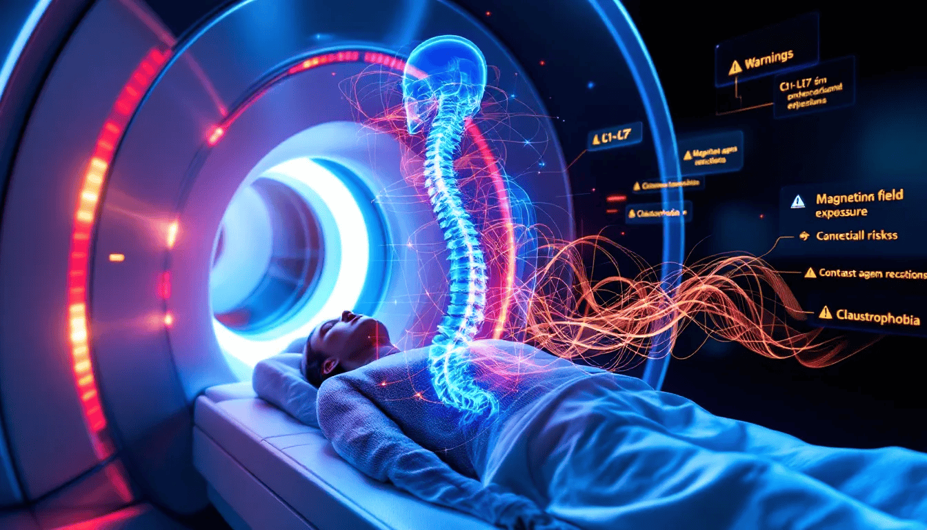
The magnetic field from an MRI is generally safe, but it can cause issues with certain implanted medical devices. For instance, patients with pacemakers or cochlear implants need careful screening to ensure their safety during the scan. Pregnant women may undergo non-contrast MRI scans if deemed necessary, although the use of contrast dyes is typically avoided.
Gadolinium contrast dye material, often used in MRI, has a lower likelihood of causing allergic reactions compared to iodine-based contrasts. However, contrast agents can still cause allergic reactions in some patients, necessitating careful screening before the procedure. Thus, while MRI scans are generally safe, specific risks must be evaluated on a case-by-case basis.
Limitations of Spine MRI Scans
While highly effective, spine MRI scans have certain limitations. Research indicates that MRI may not always accurately classify patients with lumbar disc herniation, leading to potential mismanagement. An MRI scan on its own is not sufficient for diagnosis and should be combined with clinical evaluations and other imaging techniques. Motion artifacts can significantly degrade the quality of MRI images, leading to inaccurate diagnoses.
MRI imaging is sensitive but can show abnormalities that may not correlate with clinical symptoms, leading to potential false positives. Additionally, patients with larger body sizes may not fit into standard MRI machines, limiting accessibility. MRI is also less effective in evaluating acute trauma cases compared to other imaging modalities like CT, particularly in assessing soft tissue injuries.
Cost and Accessibility of Spine MRI Scans
The starting cost of a private spine MRI scan is £249, making it an accessible option for many patients. Private spine MRI scans are available at various locations throughout the UK, ensuring easy access for patients. Vista Health, for example, offers private spine MRI scans in many locations, making them widely available.
Patients do not need a GP referral for a private spine MRI scan; they can directly access services through self-referral. To self-refer for a spine MRI scan, you need to fill in a self-referral form.
Vista Health allows patients to book online and offers flexible payment options, including spreading the payment over three months with no interest. With extended hours and appointments on weekends and evenings, scheduling a spine MRI scan has never been more convenient.
Summary
In summary, spine MRI scans are a powerful diagnostic tool that provides detailed images of the spinal column and surrounding soft tissues. They are crucial for diagnosing a wide range of conditions, from spinal infections and tumors to disc disease and nerve compression. Understanding the types, uses, and procedures involved in spine MRI scans can help patients make informed decisions about their spinal health.
While generally safe, MRI scans do come with certain risks and limitations. However, the benefits often outweigh these concerns, especially when early diagnosis and effective treatment are at stake. If you’re experiencing persistent back pain or other spinal issues, consulting with your doctor about a spine MRI scan could be a crucial step towards relief and recovery.
Frequently Asked Questions
What is a spine MRI scan?
A spine MRI scan is a noninvasive imaging test that utilizes magnetic fields and radiofrequency pulses to produce detailed images of the spinal column and surrounding soft tissues, aiding in the diagnosis of various spinal conditions without exposing the patient to radiation.
When should I consider getting a spine MRI scan?
You should consider getting a spine MRI scan if you experience severe or persistent back pain, particularly when accompanied by neurological symptoms such as numbness, tingling, or loss of bladder control. Additionally, if your pain lasts more than a few weeks and disrupts your daily activities, it is advisable to seek an MRI.
How should I prepare for a spine MRI scan?
To prepare for a spine MRI scan, inform the medical staff of any health issues, surgeries, allergies, or pregnancy, and change into a hospital gown while removing any jewelry. You typically can eat and drink beforehand unless your doctor has provided different instructions.
What are the risks associated with spine MRI scans?
Spine MRI scans are generally safe, but they carry risks for individuals with certain implanted medical devices and may cause allergic reactions to contrast agents. It is crucial to consult with your doctor about these potential risks before undergoing the procedure.
What does it mean if my spine MRI results show abnormalities?
Abnormalities in your spine MRI results may not always indicate a serious issue, as their significance depends on your symptoms and medical history. It is essential to consult a specialist for a thorough interpretation and guidance on the next steps.