Surgical management of a high grade Spondylolisthesis – The UK Spine Centre
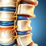
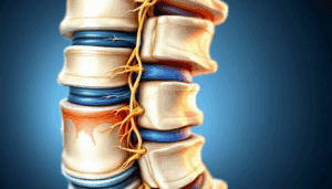
As an experienced Spinal Surgeon, I will regularly see patients with a spondylolisthesis (vertebral slippage). Most commonly this is a degenerative spondylolisthesis occurring at L4/5 due to degeneration and failure of the facet joints. Typically the slip is low grade and patients present with back and leg symptoms due to compromise of either the exiting L4 nerve root or traversing L5 nerve root under the facet joint. I also regularly see an isthmic spondylolisthesis at L5/S1 due to L5 pars defects. These slips are typically higher grade and catch the L5 root in an exiting (foraminal) location. High-grade spondylolisthesis (HGS) is diagnosed in up to 11.3% of adults with spondylolisthesis and typically presents as nonspecific lower back pain. With either of these pathologies once symptoms become established it is likely that problems will persist, worsen, or relapse with time. Both conditions can be effectively treated with surgery. Severe spondylolisthesis, including grade IV cases, present unique surgical challenges and often require specialized surgical approaches. Let me describe one such case.
JC had a congenital or dysplastic spondylolisthesis (Wiltse Classification (1976) Type I)which is seen in about 15% of cases. This is characterised by a dysplastic L5/S1 segment with hypoplastic L5/S1 facet joints, a domed sacrum, trapezoidal L5 body, L5 pars defects, L5/S1 segmental kyphosis, and a spina bifida occulta. This had resulted in a high grade slip of L5 on S1. Isthmic dysplastic spondylolisthesis and high grade dysplastic spondylolisthesis have important implications for surgical planning due to their abnormal morphology and instability.
His sagittal profile was striking but typical (Phalen-Dickson sign) for a high grade spondylolisthesis with a retroverted pelvis (anterior hip centers), increased pelvic tilt, vertical sacrum, extended hips and flexed knees. These were compensatory changes adopted during childhood and adolescence – as his slip had progressed – to maintain an upright posture. The assessment of pelvic incidence is crucial in evaluating sagittal alignment and planning surgical intervention for high grade spondylolisthesis.
His back pain and L5 radicular symptoms had deteriorated as his spondylolisthesis had progressed and he was at the limit of his compensatory changes necessary for him to be able to stand upright. Untreated the spondylolisthesis would have progressed with worsening symptoms and potentially neurological compromise leading to bilateral foot drop. Cauda equina syndrome is a critical condition that may result from high-grade spondylolisthesis, leading to loss of bladder and bowel control. In pediatric high grade and severe adolescent isthmic spondylolisthesis, early recognition and management are essential due to the risk of rapid progression and unique considerations in the growing spine.
Many different operations have been described in the last century to treat higher grade spondylolisthesis involving anterior, posterior and combined approaches to the spine. Gaines (1985) described performing an L5 vertebrectomy for the treatment of a spondyloptosis (L5 vertebra lying in front of the sacrum) bringing the L4 vertebra down onto S1. This is a long, demanding, and fairly extreme procedure which is not widely practiced. Decompression without fusion destabilises the L5 vertebra further often leading to slip progression and poor outcomes (Gill laminectomy 1984). The lumbar spine is particularly susceptible to high grade lumbosacral spondylolisthesis and high grade isthmic spondylolisthesis, which require careful surgical planning.
Surgery has two goals. Decompression to relieve neurological symptoms and stabilisation (fusion) of the unstable segment to prevent slip progression and improve back pain. Posterior decompression is especially important in high grade lumbosacral spondylolisthesis to relieve nerve root compression and reduce neurological risk.
Controversy exists between surgeons as to whether to attempt to reduce the slip or to fuse the vertebrae in situ. Advantages of reduction include normalisation of spinal alignment (allowing compensatory changes to unwind) which may reduce the chance of developing back pain from adjacent levels in the future. Also, reduction of the spondylolisthesis increases contact area between the vertebrae and increases the chance of achieving fusion. Disadvantages include increased risk of neurological deficit from stretching the nerves and higher rates of instrumentation failure. Neurological injuries are more commonly reported with reduction techniques compared to in situ fusion techniques. Longer pedicle screw constructs extending up to L4 or even L3 may be used to try and maintain reduction but a longer fusion sacrifices motion segments and may lead to accelerated degeneration at the adjacent motion segment. The debate between situ fusion techniques and reduction and fusion techniques centers on their impact on clinical and radiographic outcomes, with each approach offering distinct benefits and risks in the surgical treatment of high grade spondylolisthesis. The Meyerding classification is used to grade lumbar spondylolisthesis, with grade II and grade IV representing increasing severity of vertebral slip.
I decided with JC to attempt a partial reduction of his spondylolisthesis via a single posterior approach. I also felt it important in a young adult to avoid a long fusion construct and attempt to preserve the L4/5 motion segment if possible, reducing the chance of him developing accelerated adjacent segment degeneration at L3/4. Not fixing up to L4 however increases the risk of screw pullout, instrumentation failure, and loss of correction. In the reduction of high grade spondylolisthesis, the choice between partial and complete reduction must balance the potential for improved alignment against the risk of neurological injury.
The first stage of the operation was to expose the back of his spine across the lumbo-sacral junction. His underlying spina bifida ment the back of the dura was not covered by the bony lamina so great care was taken not to breach the dural sac (CSF leak) during the exposure. With high grade slips the L5 body lies very anteriorly and is difficult to access. The deficient L5 lamina was loose (‘rattler’) and removed. The exiting L5 roots were carefully exposed allowing direct visualisation. The L5/S1 disc space was found with the help of intraoperative X-ray imaging. Lateral radiograph is essential in evaluating slip severity and sagittal balance both preoperatively and intraoperatively. Sequential reamers were introduced into the disc space to remove disc material, flatten off the domed sacrum, and loosen up the space as far anteriorly as possible (remembering that the major vessels lie just anterior to L5) to facilitate reduction. Extra long pedicle screws were placed at S1 coming out the front of the sacrum to achieve a stronger bicortical hold. L5 pedicle screws are very challenging to place with high grade slips. Long tab screws were used at L5 allowing and an oversized rod (6.35mm), which would better withstand deformation, to be sequentially reduced. The L5 roots were visualised and probed for undue tension during the reduction stage. There was insufficient space to be able to safely introduce interbody cages into the L5/S1 interspace which would usually be my preference in a lower grade spondylolisthesis. Posterolateral fusion and interbody fusion are commonly used surgical procedures for stabilizing the lumbar spine in spondylolisthesis treated surgically. Anterior interbody fusion is also an alternative approach in selected cases.
There is always a race against time between fusion occurring and failure of the metalwork. This would have been a disaster in JC resulting in revision surgery and the construct being extended to perhaps L3. For this reason I used bone morphogenic protein which is expensive but potently and rapidly stimulates fusion. This was introduced into the L5/S1 interspace. The use of bone graft is critical in promoting successful fusion and long-term spinal stability.
Post operatively he had no neurological deficit and mobilised the day after surgery. He was only allowed to sit, lie, walk and stand over the first three months, taking great care not to unduly stress the instrumentation. At six months the L5/S1 segment had fused and he was pain free. He is now returning to all activities. Evaluating both functional and radiological outcomes is essential to assess the success of the surgical procedure and guide further management.
Very high grade slips like this are rare and I see them every few years. They present a challenging problem for the surgeon, but outcomes can be excellent if complications can be avoided. Chronic pain and functional limitations from high-grade spondylolisthesis can affect a patient’s mental health, leading to depression and anxiety. The importance of clinical outcome measures in evaluating the effectiveness of spine surgery and spinal surgery for high grade spondylolisthesis treated with various surgical procedures cannot be overstated. Spondylolisthesis treated with modern surgical techniques requires long-term follow-up, especially in high grade spondylolisthesis treated surgically. Optimal surgical intervention should be tailored to the individual patient, considering the variety of surgical approaches and surgical treatments available. The literature, including j bone joint surg, bone joint surg, spine phila pa 1976, eur spine, int j spine surg, and j spinal disord tech, provides valuable insights into surgical management and outcomes. Orthopaedic surgery is the specialty primarily involved in the surgical management of spondylolisthesis. Posterior fixation and posterior instrumentation, including pedicle screw fixation, play a key role in maintaining spinal stability, and the mobilization or removal of posterior facet joints is often necessary during surgical planning. The risk of cauda equina syndrome is a serious complication of high grade spondylolisthesis treated surgically and must be considered. Physical therapy remains important in the nonoperative management of low grade spondylolisthesis to manage symptoms and prevent progression.
Classification and Diagnosis
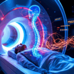
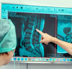
Accurate classification and diagnosis are fundamental in the management of high grade spondylolisthesis. This condition is defined by a significant forward displacement of one vertebra over another, with high grade slips typically referring to a slip percentage greater than 50%. The Meyerding classification system is widely used to grade spondylolisthesis, with grade III and above indicating high grade involvement. Clinical evaluation is complemented by advanced imaging modalities, such as magnetic resonance imaging (MRI), which provides detailed visualization of the spinal cord, nerve roots, and surrounding soft tissues. MRI is particularly valuable for assessing the degree of nerve compression and planning surgical intervention. Computed tomography (CT) scans may also be utilized to evaluate bony anatomy and the extent of vertebral displacement. Proper classification of the grade spondylolisthesis and identification of high grade slips are essential for determining the most appropriate treatment strategy and predicting clinical outcomes.
Risk Factors and Progression
Understanding the risk factors that contribute to the progression of high grade spondylolisthesis is crucial for timely intervention and optimal patient outcomes. High grade slips are more frequently observed in pediatric and adolescent populations, where the spine is still developing and more susceptible to deformity. Key risk factors include the patient’s age, the slip angle, and the presence of posterior element dysplasia, such as spina bifida occulta or elongated pars interarticularis. A higher slip angle is associated with an increased likelihood of slip progression, making it an important parameter in clinical assessment. Additionally, anatomical abnormalities in the posterior elements can compromise spinal stability and accelerate the progression of grade spondylolisthesis. Recognizing these risk factors allows clinicians to monitor patients more closely and to intervene surgically before significant neurological compromise or further slip progression occurs.
Surgical Management Options
The surgical management of high grade spondylolisthesis encompasses several approaches, each tailored to the severity of the slip and the individual patient’s needs. In situ fusion is a technique where the vertebrae are fused in their current, displaced position, which can be advantageous in minimizing neurological risk but may not correct sagittal alignment. In situ fusion techniques are often simpler and pose less risk of neurologic complications compared to reduction and fusion techniques, which better restore spino-pelvic balance. Alternatively, reduction and fusion aim to realign the vertebrae before stabilizing them, potentially restoring more normal spinal mechanics but with a higher risk of nerve injury. In the most severe cases, vertebrectomy—removal of the affected vertebra—may be considered, though this is reserved for extreme situations due to its complexity. Posterior lumbar interbody fusion (PLIF) and anterior lumbar interbody fusion (ALIF) are commonly employed surgical techniques, offering robust stabilization and the potential for improved fusion rates. The choice between these surgical techniques depends on factors such as the grade spondylolisthesis, the presence of neurological symptoms, and the patient’s overall health, with the goal of achieving both spinal stability and optimal functional outcomes.
Surgical Approach and Technique
Selecting the appropriate surgical approach and technique is vital in the treatment of high grade spondylolisthesis. The posterior approach is most commonly used, providing direct access to the posterior elements of the spine and allowing for decompression, reduction, and fusion. In some cases, an anterior approach may be employed, involving dissection through the anterior longitudinal ligament to access the intervertebral disc space and facilitate anterior lumbar interbody fusion. The surgical technique typically involves meticulous dissection to expose the affected vertebrae, followed by reduction and fusion using specialized instrumentation such as pedicle screws and rods. In certain scenarios, a transsacral interbody fusion may be performed to enhance stability, particularly in cases where traditional approaches are insufficient. The choice of surgical approach and technique is influenced by the grade spondylolisthesis, the anatomical characteristics of the patient, and the surgeon’s experience, with the overarching aim of achieving a stable, well-aligned spine and minimizing the risk of complications.
Insertion of Instrumentation
The insertion of instrumentation is a cornerstone of surgical management for high grade spondylolisthesis, providing the necessary stability to maintain spinal alignment and promote fusion. Pedicle screws are carefully placed into the pedicles of the involved vertebrae, offering strong anchorage for the attachment of rods that span the affected segment. This construct allows for controlled reduction of the slip and maintains the correction achieved during surgery. The use of high-quality, biocompatible materials such as titanium or stainless steel ensures durability and reduces the risk of hardware-related complications. Precise placement of pedicle screws is essential, particularly in high grade cases where anatomical landmarks may be distorted. Advanced imaging and intraoperative navigation can assist in achieving optimal screw trajectory and minimizing the risk of nerve injury. The successful insertion of instrumentation is critical for the long-term success of surgical management in patients with grade spondylolisthesis, supporting both immediate stability and long-term fusion.