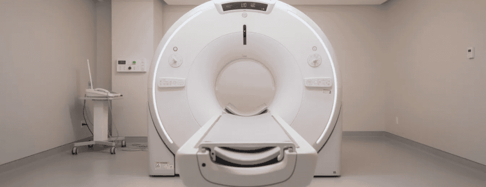
When To Get An MRI For Back Pain (From A Spinal Surgeon’s Perspective)

As a spinal surgeon, one of the most common questions I hear is “when to get an MRI for back pain.” The short answer is: most people do not need one. Let me explain when a scan genuinely helps and when it can actually do more harm than good.
Key Takeaways
- Most back pain improves within 4–6 weeks with simple measures like staying active, physiotherapy, and pain relief. An MRI for back pain is not needed immediately for most cases.
- Less than 1% of back pain cases require urgent imaging. Red flag warning signs that need same-day or next-day MRI include new leg weakness, loss of bladder or bowel control, a known history of cancer, fever, or significant trauma.
- A planned MRI is sensible when back or leg pain lasts longer than 6–12 weeks despite good treatment, or when nerve symptoms such as numbness or muscle weakness are severe and not settling.
- Unlike x rays, which show bones and arthritis but miss discs and nerves, MRI scans reveal nerves, discs, and soft tissues without any radiation exposure.
- Many scan “abnormalities” are normal age-related changes. Scans show abnormalities in 87% of pain free individuals. At the UK Spine Centre, every decision combines symptoms, examination, and imaging – never the scan alone.

Understanding Back Pain: Why Magnetic Resonance Imaging (MRI) Scans Are Not Always Needed
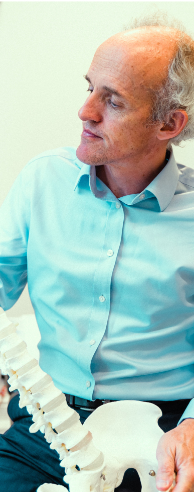
I’m Caspar Aylott, Lead Consultant Spinal Surgeon at the UK Spine Centre. I want to reassure you from the outset: back pain is extremely common, and the vast majority of people recover without any scan at all.
Doctors classify low back pain by how long it lasts: acute (under 6 weeks), subacute (6–12 weeks), and chronic (over 3 months). Research consistently shows that most acute episodes improve significantly in the first 4–6 weeks, especially with gentle movement, simple medication, and physiotherapy.
Around 85% of back pain cases are non-specific, meaning there is no single identifiable structural cause. Strained muscles, irritated joints, and age-related changes in the lumbar spine are the usual culprits, and these rarely show up neatly on any imaging test. That is why MRI is not recommended for uncomplicated low back pain initially – the first step is always a careful assessment of your medical history and symptoms by a GP, physiotherapist, or spinal specialist. Symptoms that improve with movement typically do not require an immediate MRI.
Red Flags: When Back Pain Needs An Urgent MRI Scan
While serious spinal conditions are rare, they do happen. An urgent MRI – often performed the same day – is needed when there is concern about conditions such as cauda equina syndrome, spinal infection, fracture, or cancer spread to the spine. Less than 1% of people with low back pain fall into this category, but recognising the signs can prevent permanent nerve damage.
Seek emergency help if you develop any of these red flag symptoms:
- New difficulty passing urine, or loss of bladder or bowel control
- Numbness around the bottom, genitals, or inner thighs (saddle numbness)
- Quickly worsening leg weakness or difficulty walking
- Severe back pain after a fall, car accident, or heavy injury
- Back pain with a known cancer, unexplained weight loss, or night sweats
- Back pain with fever, feeling very unwell, or signs of infection
These signs may indicate pressure on the spinal cord or nerves, or a serious underlying illness. MRI scans are used for serious spinal disorders like cancer, and MRI is essential in these urgent situations. Waiting weeks for physiotherapy alone is not appropriate here – urgent hospital review and spinal imaging are needed without delay.
When Persistent Back Pain Justifies A Planned MRI
Most MRI scans for back pain are planned, not urgent. The main indication is when pain does not improve as expected despite good treatment.
As a general guide, consider magnetic resonance imaging if:
- Low back pain or leg symptoms persist longer than 6–12 weeks
- You have had a reasonable trial of treatment – physiotherapy, regular exercise, and pain relief – without sufficient improvement
- Persistent pain after 4–6 weeks may warrant further investigation
Symptoms that commonly lead me to arrange a planned MRI include:
- Persistent sciatica or shooting pain down the leg
- Ongoing numbness, pins and needles, or weakness in a leg
- Pain that is worse in the leg than in the back, or worsens when coughing or sneezing
- Pain that keeps waking you at night for weeks despite treatment
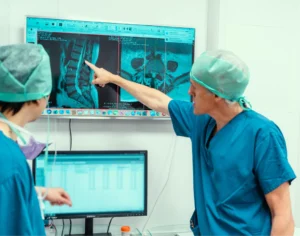
An MRI can help diagnose herniated discs, spinal stenosis, and nerve root compression – the conditions where treatment such as spinal injections or back pain surgery may genuinely help. MRI is recommended when conservative treatments fail or red flag symptoms are present.
At the UK Spine Centre, the decision is always individual. Spine surgeons like me consider your full story, examination findings, and personal priorities when assessing back pain and related symptoms – not just how long you have been suffering.
MRI vs X‑Ray: Which Scan Is Right For Back Pain?
An MRI scan and x rays give very different information, and the choice depends on what your doctor is looking for, especially when considering ageing and keeping your spine healthy.
X rays:
- Use a small dose of radiation to show bones and overall spinal alignment
- Helpful for spotting fractures, severe arthritis, or deformity such as scoliosis – x-rays are effective for detecting fractures and dislocations
- Cannot clearly show discs, nerves, muscles, or ligaments, so they often miss common causes of nerve pain
MRI scans:
- Use magnets and radio waves – no radiation at all
- Produce detailed 3D images of discs, nerves, spinal cord, joints, ligaments, and soft tissues
- Can be performed with or without contrast dye for complex cases or after previous surgery
- MRI is effective for diagnosing conditions such as infections or tumors of the spine
Unlike x rays, MRI is far more sensitive at revealing nerve compression or disc problems. An X ray might be chosen first after a fall in an older patient with a suspected compression fracture. MRI is preferred when there are nerve symptoms, suspected infection, or a need to plan pain surgery, for example when we suspect spinal stenosis causing nerve compression.
More advanced does not always mean better – sometimes an X ray is enough, and sometimes no scan is needed at all.
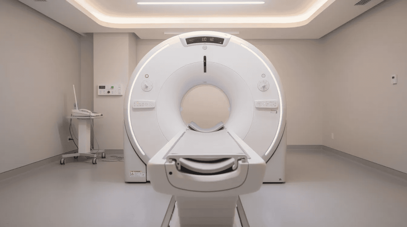
Safety Considerations: Radiation, MRI Suitability, And Contraindications
Both x rays and MRI are generally safe when used appropriately, but each has specific considerations.
Everyone receives a small amount of natural background radiation each year. A lumbar spine X ray adds a modest extra dose. Because organs in the body, including reproductive organs, are sensitive to radiation exposure, doctors avoid repeat rays unless clearly needed.
MRI involves no radiation, which is one reason it is often preferred for younger adults and when repeated imaging is necessary.
However, not everyone is suitable for an MRI:
- If you have certain metal inside your body – such as some older pacemakers, aneurysm clips, or metal fragments in the eye – MRI may not be safe
- Severe claustrophobia can make the scan difficult, though sedation or an open MRI may be discussed
- For pregnant women, most centres avoid non-urgent MRI in the first trimester unless the health benefit is clear
At the UK Spine Centre and local scanning units, every patient is carefully screened beforehand, with safety questions checked more than once to determine suitability, reflecting the same thorough approach we use across our specialised back pain diagnosis and treatment services.
What An MRI Can (And Cannot) Tell Us About Your Pain
MRI is excellent at showing the structure of your spine – what it looks like – but it cannot directly show pain. MRI does not indicate the source of back pain, and MRI does not always correlate with pain levels experienced.
Here is what surprises many patients: degenerative changes are incredibly common in people who feel perfectly fine. Disc degeneration affects 20% of 20-year-olds and 80% of 60-year-olds. Disc bulges are found in 30% of 20-year-olds and 84% of 80-year-olds. These are a normal part of ageing – almost everyone develops them.
Less than 5% of scans reveal significant issues in back pain. Age-related changes can appear on MRI scans even in symptom-free individuals, and MRI scans often show degenerative changes unrelated to pain. Some patients with severe pain have modest scan changes, and others with striking images have little or no discomfort. This is why early MRI may lead to over-diagnosis and unnecessary anxiety regarding benign findings, as echoed in many of our patient stories about spine diagnosis and treatment.
At the UK Spine Centre, every set of mri results is interpreted by a spinal specialist and carefully linked with the person’s symptoms, examination, lifestyle, and goals before recommending any spinal treatments such as injections or surgery. The relationship between what the scan shows and what you feel is not always straightforward, and that context is critical to the diagnostic process before any treatment – whether medication, injections, or surgery – is suggested. MRI findings should always be interpreted alongside symptoms and physical examination results.
How We Decide On Scans At The UK Spine Centre
Our typical pathway starts with a detailed medical history, physical and neurological examination, and review of any previous tests at our world leading London spine clinic in Harley Street. Only then do we discuss whether spinal imaging will be helpful.
Before requesting an MRI or X ray, I consider the same key questions we use in our specialist spine appointments and assessments:
- Are there any red flags or worrying features?
- Have simple treatments – activity modification, exercise, medication – been tried for a reasonable period?
- Will the scan result actually change what we do next (for example, surgery, injections, or a different type of rehabilitation)?
Since 85% of back pain cases are non-specific and do not need MRI, unnecessary scans can cause harm: radiation exposure from x rays, cost, and worry from incidental findings that are not causing your pain. Diagnosing something that does not need treatment can lead to fear and anxiety that is harder to manage than the pain itself.
When a scan is recommended, I explain the reasons in clear language, and patients are encouraged to ask questions. Some people also find it helpful to use an online back problem assessment tool such as DiagnoseMe Analyser to frame their concerns before we meet. For practical planning, private spine MRI scans in the UK typically range from around £250 to £800 depending on centre, urgency, and whether contrast is used.
Practical Advice: What To Do If You’re Unsure About Having A Scan
I understand how worrying ongoing back pain can be, and it is natural to wonder whether a scan is being missed or over-used. Neither fear nor frustration should drive the decision.
Here are simple steps you can take:
- Keep a brief symptom diary – when pain started, what worsens or eases it, any leg symptoms, and how it affects your sleep and daily life
- Book a review with your GP, physiotherapist, or spinal specialist to go through this information
- Ask directly: “Do I need an MRI or X ray now, and if not, what signs should I watch for?”
If any red flag symptoms develop after an initial assessment – even if you were told a scan was not needed – seek urgent help straight away.
If you remain uncertain, a second opinion with a spinal specialist — including a video consultation with a spine expert if travel is difficult — can provide clarity on whether imaging is appropriate. The goal is always the same: the right scan, at the right time, for the right reason. Many people recover fully without ever needing an MRI scan, and gentle guided movement is usually far more helpful than any set of images on a screen.

FAQs
Is it dangerous to delay an MRI for back pain?
In the absence of red flag symptoms, delaying an MRI for several weeks while trying conservative treatment is very unlikely to cause harm. Serious spinal conditions are rare and usually produce clear warning signs. If new bladder, bowel, or leg weakness symptoms appear at any stage, seek urgent review – but for the majority of people suffering from back pain, patience and active treatment are the safest approach.
Can I have an MRI if I already had X rays?
Yes. Having had an X ray does not stop you from having an MRI. The two tests show different things – X ray for bone structure and alignment, MRI for nerves, discs, and soft tissues. Your specialist will avoid repeating tests that do not add new information, but in many cases both are helpful at different stages of your diagnosis.
Will an MRI show exactly where my pain is coming from?
MRI often shows likely pain sources, such as a disc pressing on a nerve, but it cannot measure pain itself. MRI is not useful for diagnosing the source of back pain in isolation. At the UK Spine Centre, decisions are always based on the combination of symptoms, examination, and MRI – never the scan alone.
How should I prepare for a spine MRI scan?
Bring a list of your medications and arrive early to complete safety forms. Wear clothing without metal fastenings. Mention any implants, previous operations, or if you have any metal inside your body. The scan itself is painless, though it can be noisy. It typically takes 20–40 minutes for the lower back. You do not need to worry – staff will explain everything before you go in.
Can exercise make my back worse if I have not had a scan yet?
Gentle, guided activity is usually helpful, not harmful, for most episodes of low back pain. Follow advice from a physiotherapist or clinician, avoid heavy lifting or activities that sharply worsen symptoms, and seek reassessment if pain is rapidly getting worse or new leg symptoms or red flags appear. Staying active is one of the most effective ways to recover and is a normal part of the healing process.